Technical Specifications
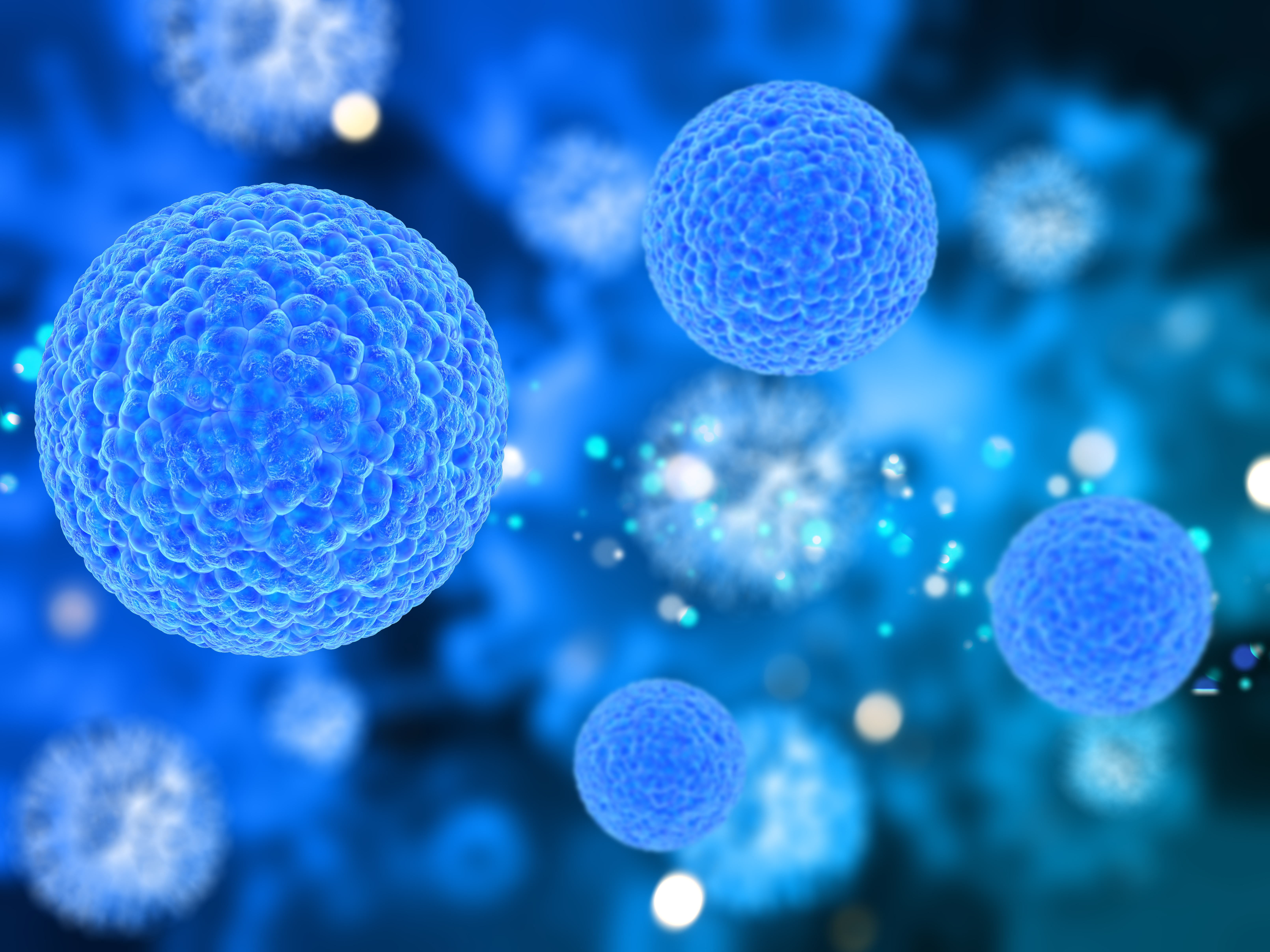
Decoding Cancer Complexity with an Integrated Solution
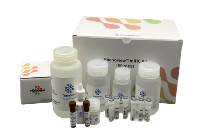
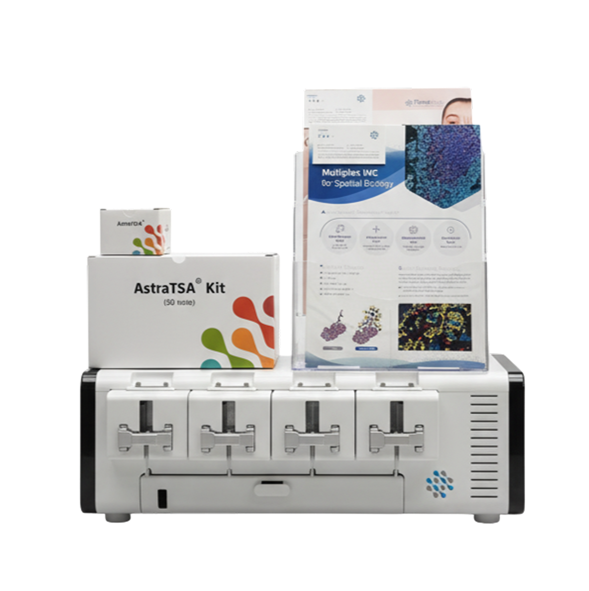
AstraTSA kit
(A high-performance, versatile Multiplex IHC product)
AstraBCF kit
(A next-generation technology enabling simultaneous imaging of proteins and RNA, optimized for drug development and biomarker discovery (Hyperplex IF))
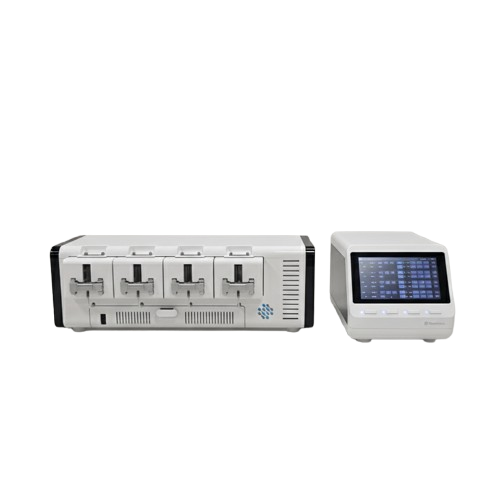
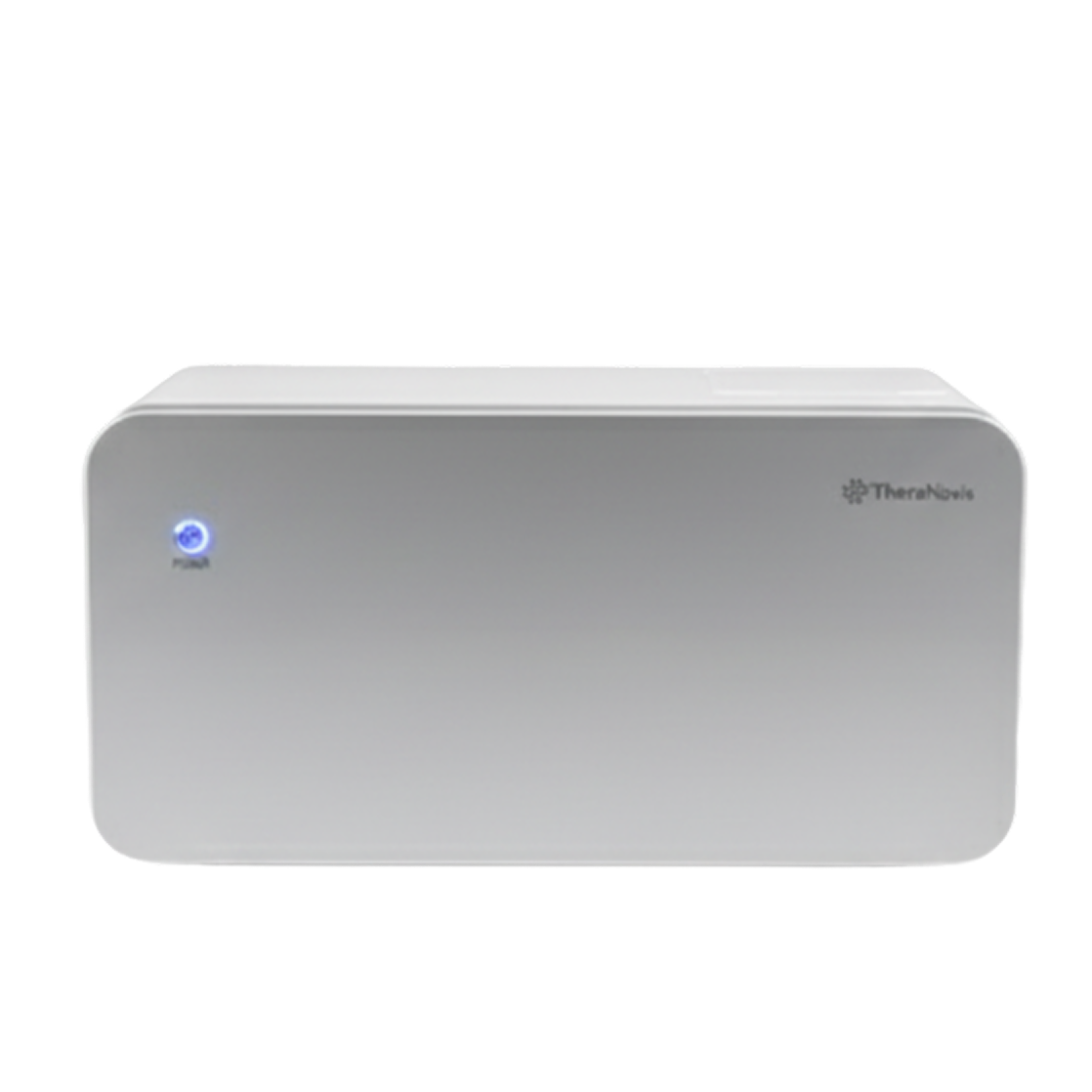
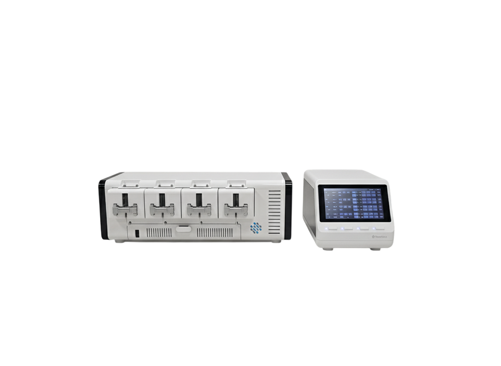
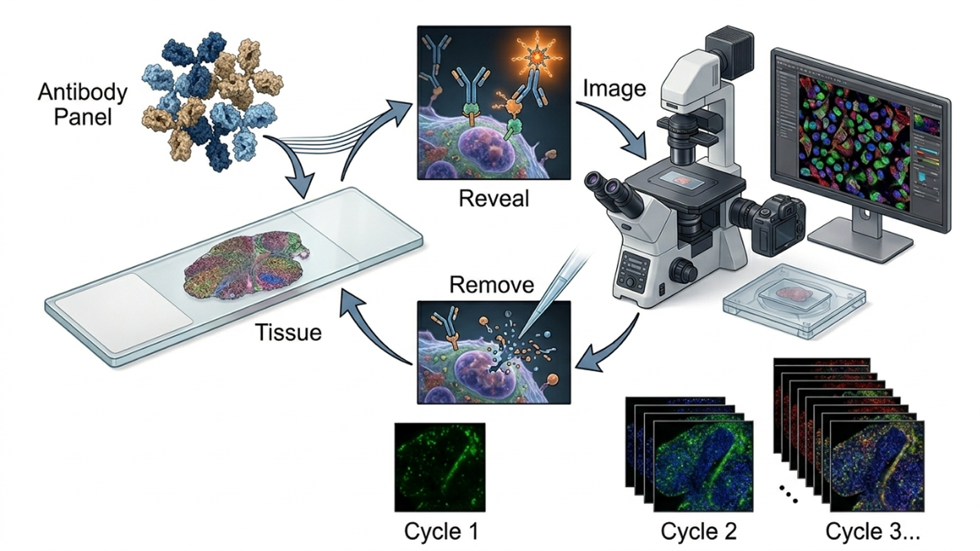
NovaStain
(an automated staining system designed to save researchers' time and improve result reliability)
Featured Product Lineup
Staining and Analysis Solutions for Spatial Proteomics
Advanced Multiplex Staining Kits Powered by Proprietary Technology
Next-generation technology enabling simultaneous protein and RNA imaging, optimized for drug discovery and biomarker identification (Hyperplex IF)
* Unprecedented level of data can be obtained from a single tissue slide at a competitive cost.
AstraBCF (up to 120-plex)
Unlock Unprecedented Data with TSA-Based Technology
Achieve high-sensitivity, multiplex staining and extract deeper insights from a single tissue slide—efficiently and cost-effectively.
Standard detection
AstraTSA
- 10-100x improvement in fluorescence signal sensitivity
- Minimizes autofluorescence
- Reduces antibody (Ab) usage to one-fifth of conventional levels
- Enables simultaneous use of multiple markers
- Compatible with signal amplification across all immunoassays
- Supports both chromogenic and fluorescent labeled probes
TSA-Based Kit
- Following the development of TSA dyes, 12 spectrally distinct fluorescent TSA dyes were produced.
- Commercialized staining kits configured in 4-plex, 7-plex, 9-plex, and 12-plex formats using fully validated panels.
- Achieved over 70% in-house integration through proprietary reagent production.
- Designed for broad compatibility, enabling easy adoption in laboratories equipped with fluorescence microscopes.
- Developed autofluorescence removal kits and preprocessing kits for skin cancer fluorescence staining.

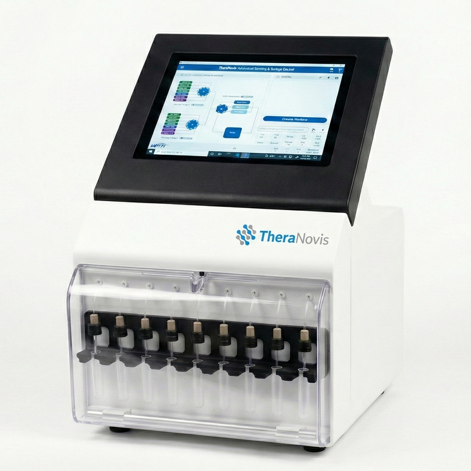
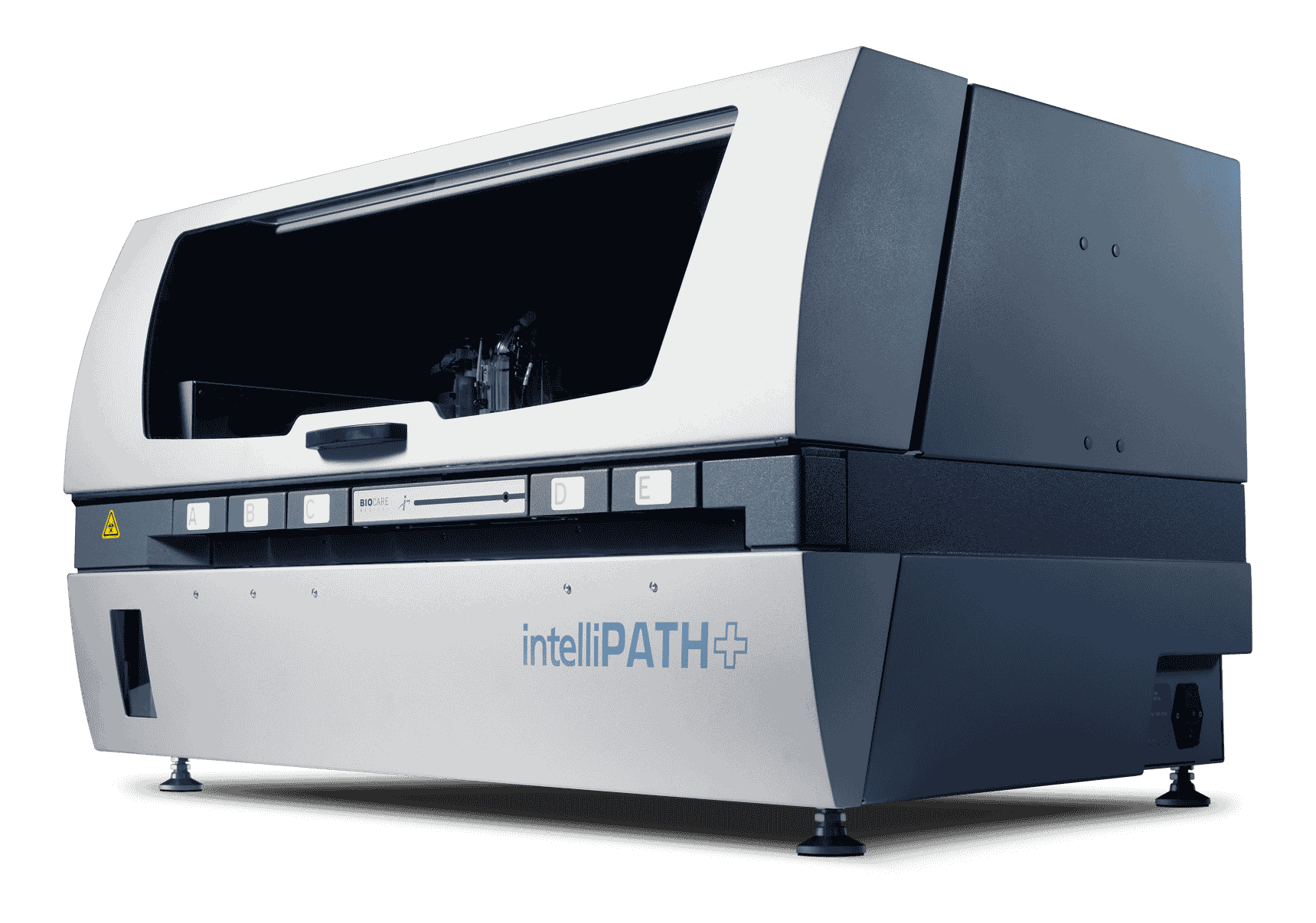
An automated staining system (NovaStain™) that saves researchers' time and enhances result reliability
AstraNavi provides an automated tissue staining system optimized for TSA-MIF, based on continuous user feedback.
Temperature Control
Precise thermal management for optimal staining
Fluidic Control
Automated reagent delivery and circulation
User Environment
Optimized workflow for laboratory efficiency
Eliminating Key Workflow Bottlenecks, Achieving Complete Standardization of Staining
Challenges1. Operator Dependency
Variability in results increases based on user skill level
2. Lack of Standardization
High variability across laboratories and operators
3. High Failure Rates and Costs
Loss of valuable samples and expensive reagents
* Standardizing the staining process determines up to 50% of overall data quality.
By controlling variables in the staining process, we ensure reliable data across the entire workflow
Our Solution1. Automated Reproducibility
Automation of multiplex staining processes eliminates user-to-user variability
2. Software-Driven Quality Control
Ensures consistency through real-time process control and monitoring
3. End-to-End Integration
Enhances overall quality through seamless connection with downstream analysis
Compared to competitors, delivers higher detection sensitivity, faster processing time, and multiplexing capability—while maintaining strong cost competitiveness
| Product | Cell DIVE | CODEX | Hyperion | GeoMx | AstraNavi |
|---|---|---|---|---|---|
| Manufacturer | Cytiva | Akoya | Fluidigm | NanoString | TheraNovis |
| Number of Biomarkers | 60 | 100 | 37 | 4 | 120 |
| Channels | 5 | 4 | 135 | 4 | 12 markers / 4 channels |
| Dye | DAPI, Cy2/3/5/7 | DNA Barcoding Ab | Ab labeled with metal ions | Oligo Barcoding Ab | 12 dyes for antibodies |
| Validated Antibodies | 350+ validated Ab | 160 | >100 | 96 | 200 |
| Spatial Context | Yes | Yes | Yes | No | Yes |
| Throughput | 8 plex 1 slide/day | 35 plex 1 slide/day | 37 plex/day | 4 plex 16 slides/day | 12-plex: 1 slide / 2h, 120-plex: 1 slide / day |
Competitive Technology Comparison (Global & Domestic)
Expert-level spatial analysis reports generated in just a few clicks
Cloud-Based
Access and analyze data anytime, anywhere
Automated Analysis
Automated TIPS & CPS scoring and spatial cellular network analysis
User-Friendly Dashboard
Intuitive UI/UX with automated report generation
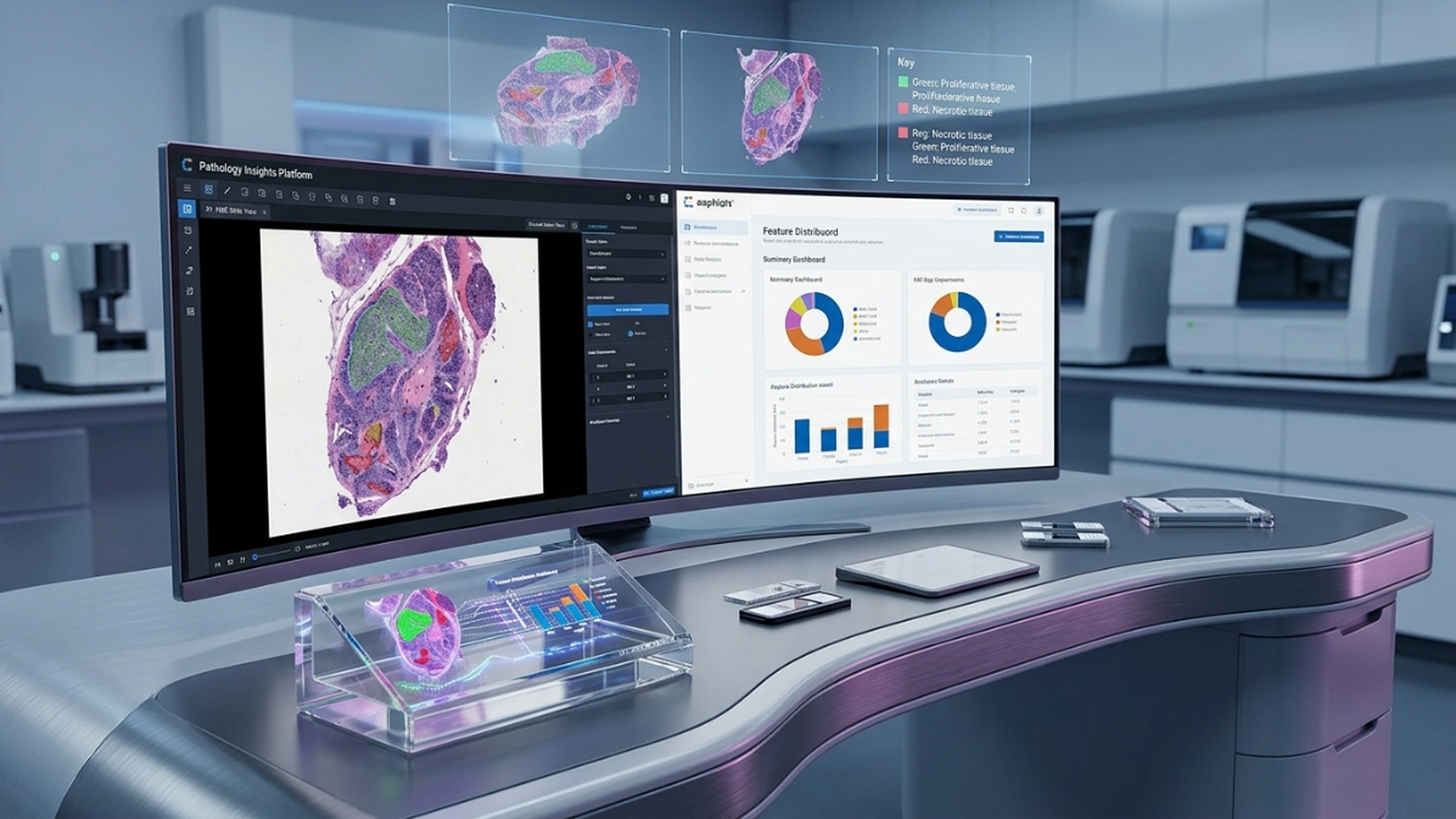
CHIEF™ Image viewer and data extraction preprocessing software
AnyPath™ Cloud-based automated analysis and reporting platform
AI-Powered Automated Analysis Software
1. Development of machine learning-based analysis technologies for multiplex IHC results
- Provides solutions that streamline image analysis workflows for digital pathology, and develops AI-driven platforms, 'CHIEF™ & AnyPath™' for fast and accurate image evaluation
- Development of systems enabling automated tumor region detection and analysis through collaboration with AI companies
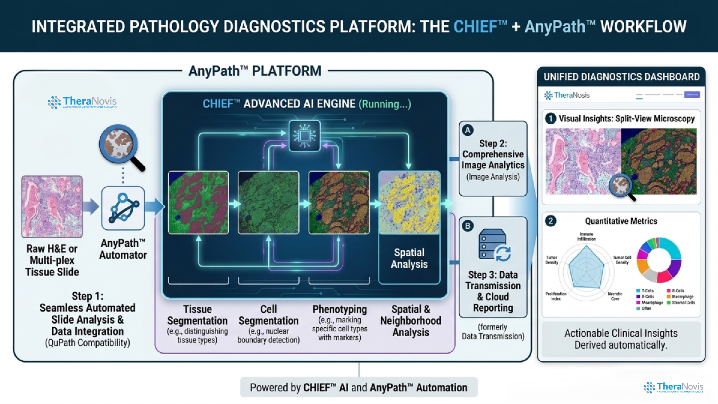
2. Development of automated TPS/CPS analysis methods (AnyPath™ PD-L1)
- Korean patent granted, PCT application filed, and U.S. patent application submitted
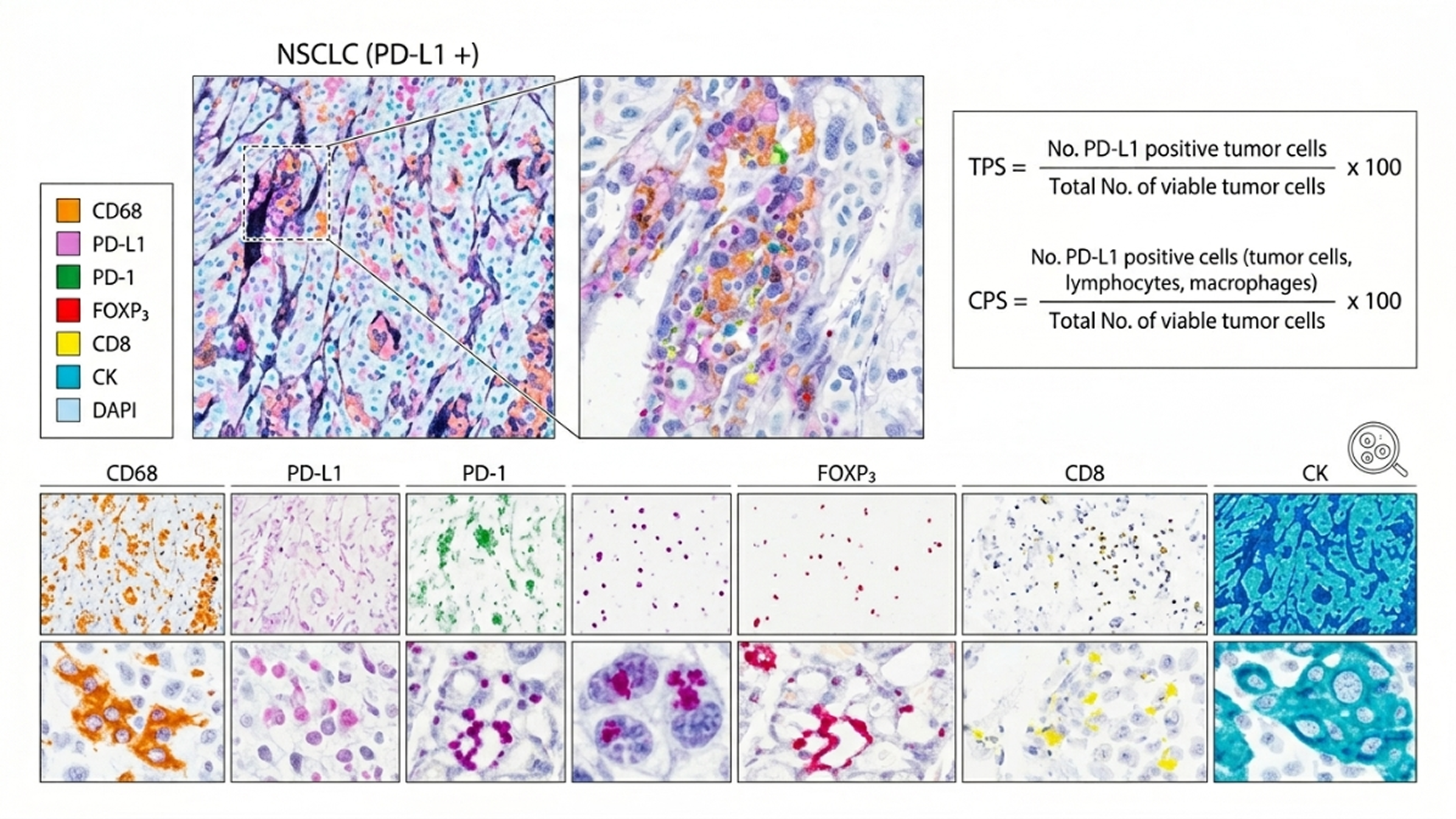
Multiplex Image Analysis Result (6-plex, 120-plex)

Discover the hidden value of your tissue samples with our Advanced Spatial Proteomics Solution.
Join leading researchers in unlocking the next frontier of precision medicine.